Back to Profile Page

Tree of Life Media Contributed By Miroslav Hylis

<< < 1 2 3 4 > >>
ID Thumbnail Media Data
30987
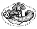
Scientific Name Metchnikovella sp.
Comments N- nucleus, F- polar filament, t- tubules
Reference Weiser J. 1977. Contribution to the classification of microsporidia. Věstník Československé spolecnosti zoologické 41 (4): 308-312.
Acknowledgements reproduced with permission
Life Cycle Stage spore
Copyright © 1977 Českoslovensé zoologické společnosti
Image Use ToL use only
Attached to Group Metchnikovella (Microsporidia): view page image collection
Title metchkresba.jpg
Image Type Diagram
Image Content Specimen(s), Ultrastructure
Technical Information schematic representation of spore structure
ID 30987
30988
Scientific Name Chytridiopsis-like microsporidium
Comments Chytridiopsis-like microsporidium from Biomphalaria sp. erronerously identified as a Coccospora sp.
Specimen Condition Dead Specimen
Life Cycle Stage spore
Copyright © 1969 C. S. Richards
Image Use ToL use only
Attached to Group Chytridiopsis-like microsporidia (Microsporidia): view page image collection
Title coccospora2a.jpg
Image Type Photograph
Image Content Specimen(s), Ultrastructure
Technical Information TEM micrograph
ID 30988
30989
Scientific Name Chytridiopsis sp.
Comments schematic representation of spore structure
Reference Canning E.U., Vavra J. 2000. Phylum Microsporida Balbiani, 1882. Pp. 39-126. In: An Illustrated Guide to the Protozoa. Second Edition. Vol. 1. (Lee J. J., Leedale G.F. and Badbury P., eds.). Society of Protozoologists, Lawrence, Kansas, U.S.A.
Acknowledgements used with permission
Life Cycle Stage spore
Body Part showing anchoring disk and honeycomb polar tube
Copyright © 2000 Society of Protozoologists
Image Use ToL use only
Attached to Group Chytridiopsis-like microsporidia (Microsporidia): view page image collection
Title chytrdillguide2.jpg
Image Type Diagram
Image Content Specimen(s), Ultrastructure
ID 30989
30158
Scientific Name Binucleata daphniae
Comments spores(about 5.0 x 2.5 μm in size) isolated from infected hypodermal cells of infected Daphnia. Each spore shows a vacuole-like area in the posterior pole of the spore. This is a specific character of microsporidian spores.
Specimen Condition Live Specimen
Life Cycle Stage spores
Copyright © 2008
Image Use restricted
Attached to Group Binucleata (Microsporidia): view page image collection
Title binucleatasporynom2.jpg
Image Type Photograph
Image Content Specimen(s)
Technical Information Nomarski phase contrast micrograph
ID 30158
30161
Comments Spore of Edhazardia aedis, parasite of the mosquito Aedes aegypti with fully everted polar tube showing the length of the filament in comparison with spore size (about 8 μm).
Specimen Condition Live Specimen
Life Cycle Stage spore
Copyright © 2005 J.J. Becnel
Image Use ToL use only
Attached to Group Microsporidia: view page image collection
Title sporavystrel-lm.jpg
Image Type Photograph
Image Content Specimen(s), Body Parts
ID 30161
30163
Scientific Name Nosema tractabile
Specimen Condition Dead Specimen
Life Cycle Stage spores
Copyright © 2004 Ronny Larsson
Image Use ToL use only
Attached to Group typical microsporidia (Microsporidia): view page image collection
Title sporavystrelsem.jpg
Image Type Photograph
Image Content Body Parts, Ultrastructure
Technical Information scanning electron micrograph
ID 30163
30165
Comments Generalized microsporidian life cycle. (a) the dormant spore containing a coiled polar filament. (b) upon germination the polar filament everts from the spore as a tube which pierces the host cell membrane. (c) the spore content in the form of a small, little organized cell (sporoplasm) is deposited in the cytoplasm of the host cell. (d) the injected sporoplasm reorganizes its cytoplasm and nucleus, becomes a meront, and starts the vegetative part of the life cycle, the merogony. (e) in sporogony cells of the parasite acquire cell walls and gradually change into spores.
Reference Keeling, P. J. and G. I. McFadden. 1998. Origins of microsporidia. Trends Microbiol. 6:19-23.
Copyright © 1998 Elsevier Ltd.
Image Use ToL use only
Attached to Group Microsporidia: view page image collection
Title zivcyklschema.jpg
Image Type Diagram
Notes permission pending
ID 30165
30166
Reference Vemuganti, G. K., P. Garg, S. Sharmal, J. Joseph, U. Gopinathan, and S. Singh. 2005. Is Microsporidial keratitis an emerging cause of stromal keratitis? – a case series study. BMC Ophthalmology 2005, 5:19 doi:10.1186/1471-2415-5-19
Specimen Condition Live Specimen
Body Part cornea
Source Is Microsporidial keratitis an emerging cause of stromal keratitis? – a case series study
Image Use creative commons This media file is licensed under the Creative Commons Attribution License - Version 2.0.
Copyright © 2005 Vemuganti et al.
Attached to Group Homo sapiens: view page image collection
Microsporidia: view page image collection
Title oko-keratitis.jpg
Image Type Photograph
Image Content Body Parts
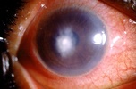
Technical Information Slit lamp biomicroscopy of cornea in diffuse illumination with central mid to deep stromal infiltrate and surrounding stromal edema.
ID 30166
30167
Reference Vemuganti, G. K., P. Garg, S. Sharmal, J. Joseph, U. Gopinathan, and S. Singh. 2005. Is Microsporidial keratitis an emerging cause of stromal keratitis? – a case series study. BMC Ophthalmology 2005, 5:19 doi:10.1186/1471-2415-5-19
Specimen Condition Dead Specimen
Source Is Microsporidial keratitis an emerging cause of stromal keratitis? – a case series study
Image Use creative commons This media file is licensed under the Creative Commons Attribution License - Version 2.0.
Copyright © 2005 Vemuganti et al.
Attached to Group Microsporidia: view page image collection
Title keratitislm21.jpg
Image Type Photograph
Image Content Specimen(s), Body Parts
Technical Information Section of corneal button in which the spores appear red with darkly stained band at the tip (1% acid-fast stain, ×1000).
ID 30167
30986
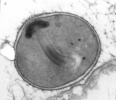
Scientific Name Metchnikovella sp.
Specimen Condition Dead Specimen
Life Cycle Stage spore
Copyright © 1973 Joseph Schrevel
Image Use ToL use only
Attached to Group Metchnikovella (Microsporidia): view page image collection
Title metchnikpas.jpg
Image Type Photograph
Image Content Specimen(s), Ultrastructure
Technical Information TEM micrograph of PAS stained specimen
ID 30986
Please note: Most images and other media displayed on the Tree of Life web site are protected by copyright, and the ToL cannot act as an agent for their distribution. If you would like to use any of these materials for your own projects, you need to ask the copyright owner(s) for permission. For additional information, please refer to the ToL Copyright Policies.

Contribute to the ToL

ToL Contributions

Ways to Contribute

Use of Contributions

Scientific Content

Building Treehouses

Images/Media

  go to the Tree of Life home page
top